プロテアーゼ活性化受容体1
プロテアーゼ活性化受容体1(プロテアーゼかっせいかじゅようたい1、英: protease-activated receptor 1、略称: PAR1)は、ヒトではF2R遺伝子によってコードされるタンパク質である[5]。トロンビン(凝固第II因子)受容体という語がこのタンパク質を指して用いられることもある。PAR1はGタンパク質共役受容体であり、4種類のプロテアーゼ活性化受容体の1つである。血小板と内皮細胞で高度に発現しており、血液凝固と炎症の連携を媒介する重要な役割を果たし、炎症性肺疾患や線維性肺疾患の発症に重要である[6]。また、トロンビンまたは活性化プロテインCとの相互作用を介して、血管内皮のバリアの完全性の破壊と維持にそれぞれ関与している[7]。




構造
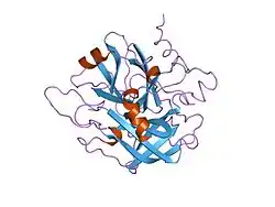
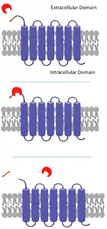
PAR1は膜貫通Gタンパク質共役受容体(GPCR)であり、425アミノ酸残基からなる。その構造の特徴は他のプロテアーゼ活性化受容体と多くが共通しており[8][9]、7本の膜貫通αヘリックスを持ち、3つの細胞外ループと3つの細胞内ループを持つ[9]。N末端へは細胞外に位置し、トロンビンの結合に適した配置をしている。C末端は細胞内側に位置し、細胞質テールの一部をなしている[8]。
シグナル伝達経路
活性化
PAR1はN末端の41アミノ酸がトロンビンによって切断されることで活性化される[10]。トロンビンはN末端のLys-Asp-Pro-Arg-Ser配列を認識し、Arg41とSer42の間のペプチド結合を切断する。PAR1の特異的切断部位に対するトロンビンの親和性は、トロンビンのexositeと呼ばれる領域とPAR1のSer42のC末端側に位置するアミノ酸からなる酸性領域との二次的相互作用によってさらに強化される[11]。このタンパク質分解による切断は不可逆的で、切断されたペプチド(parstatinとも呼ばれる)はその後、細胞外へ放出される[10]。切断によって生じた新たなN末端は、PAR1の2つ目の細胞外ループの結合領域に結合するテザードリガンド(係留リガンド)として作用し、PAR1を活性化する。この結合はタンパク質のコンフォメーション変化を促進し、PAR1の細胞内領域へのGタンパク質の結合を可能にする[12][13]。
シグナル伝達
PAR1は切断を受けると、細胞内ループのいくつかの部位に結合したGタンパク質を活性化する。例えば、PAR1はPAR4とともに、G12/13型Gタンパク質と共役して活性化し、RhoとRhoキナーゼを活性化する[8]。この経路はアクチンの収縮による血小板の形状の迅速な変化を引き起こして血小板に可動性をもたらすとともに、顆粒の放出を引き起こす。どちらも血小板の凝集に必要な過程である[8]。
さらに、PAR1とPAR4の双方がGqと共役し、細胞内のカルシウムイオンの移動を刺激する。カルシウムは血小板活性化のセカンドメッセンジャーとして機能する[8]。また、この経路はプロテインキナーゼCも活性化し、血小板の凝集を促進し、血液凝固経路をさらに進行させる[11]。
終結
PAR1の細胞質テールのリン酸化とその後のアレスチンの結合は、PAR1をGタンパク質シグナル伝達から脱共役させる[10][11]。リン酸化されたPAR1はエンドソームを介して細胞内へ送り返され、ゴルジ体へ送られる。その後、切断されたPAR1は選別されてリソソームへ輸送され、分解される[11]。このインターナリゼーションと分解は、受容体シグナル伝達を終結させるために必要な過程である[10]。
細胞がトロンビンに対する応答性を再獲得するためには、PAR1が細胞膜へ再補充されなければならない。細胞膜の未切断のPAR1には、細胞内のC末端のチロシンモチーフにAP2アダプター複合体が結合し、エンドサイトーシスが促進される[14]。これらはその後、細胞質のクラスリン被覆小胞に貯蔵され、タンパク質分解から保護される。未切断のPAR1は再合成に依存しない形でこうした小胞から細胞膜へ定常的に供給され、細胞は再びトロンビンに対して感作状態となってシグナル伝達経路が再設定される[15]。
リガンド
アゴニスト
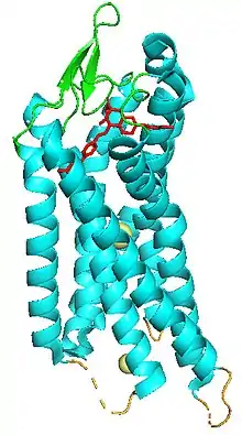
PAR1に対する選択的アゴニストの探索は、研究者の関心事となっている。合成SFLLRNペプチドはPAR1のアゴニストとして作用することが知られている。SFLLRNペプチドは活性化PAR-1のN末端テザードリガンドの最初の6残基を模倣し、2つ目の細胞外ループ上の同じ結合部位に結合する[16]。そのため、トロンビンが存在しない場合でも、SFLLRNの結合によってPAR1の切断に伴う応答をもたらすことができる[17]。
アンタゴニスト
PAR1の選択的アンタゴニストは抗凝固薬として開発されている。
- SCH-79797
- ボラパキサルはZontivityの商品名で販売されており、心筋梗塞や末梢動脈疾患の病歴がある患者の心疾患の治療に利用される、ファースト・イン・クラス(画期的医薬品)抗血小板薬である[18]。ボラパキサルは近年、IL-1βなどの炎症性サイトカインやCXCL1、CCL2、CCL7などのケモカインのレベルを低下させることにより、肺炎球菌Streptococcus pneumoniaeに対する好中球の炎症応答を弱めることが明らかにされた[19]。ボラパキサルはPAR1の細胞外ループ2と3の間の結合ポケットに結合することで阻害を行う。ボラパキサルの結合はPAR1の不活性構造を安定化し、活性化型コンフォメーションへの切り替えを防ぐ[16]。
出典
- GRCh38: Ensembl release 89: ENSG00000181104 - Ensembl, May 2017
- GRCm38: Ensembl release 89: ENSMUSG00000048376 - Ensembl, May 2017
- Human PubMed Reference:
- Mouse PubMed Reference:
- “Chromosomal assignment of the human thrombin receptor gene: localization to region q13 of chromosome 5”. Blood 82 (5): 1532–7. (September 1993). doi:10.1182/blood.V82.5.1532.1532. PMID 8395910.
- "“Proteinase-activated receptors in fibroproliferative lung disease”. Thorax 69 (2): 190–2. (February 2014). doi:10.1136/thoraxjnl-2013-204367. PMID 24186921.
- “Endothelial barrier protection by activated protein C through PAR1-dependent sphingosine 1-phosphate receptor-1 crossactivation”. Blood 105 (8): 3178–84. (April 2005). doi:10.1182/blood-2004-10-3985. PMID 15626732.
- Michelson, Alan D. (2013). Platelets (3rd ed.). Amsterdam: Elsevier. ISBN 9780123878380. OCLC 820818942
- “Structural Properties of the Human Protease-Activated Receptor 1 Changing by a Strong Antagonist”. Structure 26 (6): 829–838.e4. (June 2018). doi:10.1016/j.str.2018.03.020. PMID 29731231.
- “Signal transduction by protease-activated receptors”. British Journal of Pharmacology 160 (2): 191–203. (May 2010). doi:10.1111/j.1476-5381.2010.00705.x. PMC 2874842. PMID 20423334.
- “Protease-activated receptor signalling, endocytic sorting and dysregulation in cancer”. Journal of Cell Science 120 (Pt 6): 921–8. (March 2007). doi:10.1242/jcs.03409. PMID 17344429.
- “Identifying and quantifying two ligand-binding sites while imaging native human membrane receptors by AFM”. Nature Communications 6 (1): 8857. (November 2015). doi:10.1038/ncomms9857. PMC 4660198. PMID 26561004.
- Ramachandran, Rithwik; Noorbakhsh, Farshid; Defea, Kathryn; Hollenberg, Morley D. (2012-01-03). “Targeting proteinase-activated receptors: therapeutic potential and challenges”. Nature Reviews. Drug Discovery 11 (1): 69–86. doi:10.1038/nrd3615. ISSN 1474-1784. PMID 22212680.
- “Regulation of protease-activated receptor 1 signaling by the adaptor protein complex 2 and R4 subfamily of regulator of G protein signaling proteins”. The Journal of Biological Chemistry 289 (3): 1580–91. (January 2014). doi:10.1074/jbc.m113.528273. PMC 3894338. PMID 24297163.
- “Clathrin adaptor AP2 regulates thrombin receptor constitutive internalization and endothelial cell resensitization”. Molecular and Cellular Biology 26 (8): 3231–42. (April 2006). doi:10.1128/MCB.26.8.3231-3242.2006. PMC 1446942. PMID 16581796.
- “High-resolution crystal structure of human protease-activated receptor 1”. Nature 492 (7429): 387–92. (December 2012). doi:10.1038/nature11701. PMC 3531875. PMID 23222541.
- “Protease-activated receptor-1 can mediate responses to SFLLRN in thrombin-desensitized cells: evidence for a novel mechanism for preventing or terminating signaling by PAR1's tethered ligand”. Biochemistry 38 (8): 2486–93. (February 1999). doi:10.1021/bi982527i. PMID 10029543.
- “Vorapaxar: The Current Role and Future Directions of a Novel Protease-Activated Receptor Antagonist for Risk Reduction in Atherosclerotic Disease”. Drugs in R&D 17 (1): 65–72. (March 2017). doi:10.1007/s40268-016-0158-4. PMC 5318326. PMID 28063023.
- “Regulation of neutrophilic inflammation by proteinase-activated receptor 1 during bacterial pulmonary infection”. Journal of Immunology 194 (12): 6024–34. (June 2015). doi:10.4049/jimmunol.1500124. PMC 4456635. PMID 25948816.
関連文献
- “Characterization of a functional thrombin receptor. Issues and opportunities”. The Journal of Clinical Investigation 89 (2): 351–5. (February 1992). doi:10.1172/JCI115592. PMC 442859. PMID 1310691.
- “Time course of upregulation of inflammatory mediators in the hemorrhagic brain in rats: correlation with brain edema”. Neurochemistry International 57 (3): 248–53. (October 2010). doi:10.1016/j.neuint.2010.06.002. PMC 2910823. PMID 20541575.
- “Role of thrombin and its major cellular receptor, protease-activated receptor-1, in pulmonary fibrosis”. Biochemical Society Transactions 30 (2): 211–6. (April 2002). doi:10.1042/BST0300211. PMID 12023853.
- “Role and regulation of the thrombin receptor (PAR-1) in human melanoma”. Oncogene 22 (20): 3130–7. (May 2003). doi:10.1038/sj.onc.1206453. PMID 12789289.
- “PGE2 and PAR-1 in pulmonary fibrosis: a case of biting the hand that feeds you?”. American Journal of Physiology. Lung Cellular and Molecular Physiology 288 (5): L789-92. (May 2005). doi:10.1152/ajplung.00016.2005. PMID 15821019.
- “Protease-activated receptors in cardiovascular diseases”. Circulation 114 (10): 1070–7. (September 2006). doi:10.1161/CIRCULATIONAHA.105.574830. PMID 16952995.
- “Protease-activated receptor signaling: new roles and regulatory mechanisms”. Current Opinion in Hematology 14 (3): 230–5. (May 2007). doi:10.1097/MOH.0b013e3280dce568. PMID 17414212.
- “Molecular cloning of a functional thrombin receptor reveals a novel proteolytic mechanism of receptor activation”. Cell 64 (6): 1057–68. (March 1991). doi:10.1016/0092-8674(91)90261-V. PMID 1672265.
- “Solid tumor cells express functional "tethered ligand" thrombin receptor”. Cancer Research 55 (3): 698–704. (February 1995). PMID 7834643.
- “Intracellular targeting and trafficking of thrombin receptors. A novel mechanism for resensitization of a G protein-coupled receptor”. The Journal of Biological Chemistry 269 (44): 27719–26. (November 1994). PMID 7961693.
- “Crystallographic structures of thrombin complexed with thrombin receptor peptides: existence of expected and novel binding modes”. Biochemistry 33 (11): 3266–79. (March 1994). doi:10.1021/bi00177a018. PMID 8136362.
- “G proteins of the G12 family are activated via thromboxane A2 and thrombin receptors in human platelets”. Proceedings of the National Academy of Sciences of the United States of America 91 (2): 504–8. (January 1994). doi:10.1073/pnas.91.2.504. PMC 42977. PMID 8290554.
- “Response of blood leukocytes to thrombin receptor peptides”. Journal of Leukocyte Biology 54 (2): 145–51. (August 1993). doi:10.1002/jlb.54.2.145. PMID 8395550.
- “Genomic cloning and characterization of the human thrombin receptor gene. Structural similarity to the proteinase activated receptor-2 gene”. The Journal of Biological Chemistry 271 (16): 9307–12. (April 1996). doi:10.1074/jbc.271.16.9809. PMID 8621593.
- “Cloning and identification of regulatory sequences of the human thrombin receptor gene”. The Journal of Biological Chemistry 271 (42): 26320–8. (October 1996). doi:10.1074/jbc.271.42.26320. PMID 8824285.
- “Role of the thrombin receptor's cytoplasmic tail in intracellular trafficking. Distinct determinants for agonist-triggered versus tonic internalization and intracellular localization”. The Journal of Biological Chemistry 271 (51): 32874–80. (December 1996). doi:10.1074/jbc.271.51.32874. PMID 8955127.
- “Direct evidence for two distinct G proteins coupling with thrombin receptors in human neuroblastoma SH-EP cells”. European Journal of Pharmacology 316 (1): 105–9. (November 1996). doi:10.1016/S0014-2999(96)00653-X. PMID 8982657.
- “Thrombin receptors on human platelets. Initial localization and subsequent redistribution during platelet activation”. The Journal of Biological Chemistry 272 (9): 6011–7. (February 1997). doi:10.1074/jbc.272.9.6011. PMID 9038223.
- “Specific inhibition of thrombin-induced cell activation by the neutrophil proteinases elastase, cathepsin G, and proteinase 3: evidence for distinct cleavage sites within the aminoterminal domain of the thrombin receptor”. Blood 89 (6): 1944–53. (March 1997). doi:10.1182/blood.V89.6.1944. PMID 9058715.