尿膜
機能
この袋状の構造は消化器系および泌尿器系に接続され、血管網で覆われている。尿膜の機能は胚から液状の老廃物を集めるとともに、胚により使用されたガスを交換することである。
爬虫類、鳥類、およびカモノハシ
この構造は初め、爬虫類において窒素性の老廃物を貯蔵するとともに、胚に酸素を供給するため形成された。酸素は卵殻を介して尿膜に吸収される。卵生の哺乳類であるカモノハシ目でも、尿膜の機能は同様である。
追加画像
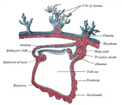 Section through the embryo.
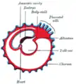 Diagram showing later stage of allantoic development with commencing constriction of the yolk-sac.
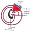 Diagram showing the expansion of amnion and delimitation of the umbilicus.
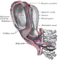 Model of human embryo 1.3 mm. long.
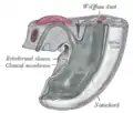 Tail end of human embryo from fifteen to eighteen days old.
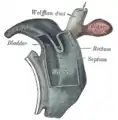 Cloaca of human embryo from twenty-five to twenty-seven days old.
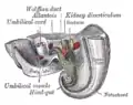 Tail end of human embryo twenty-five to twenty-nine days old.
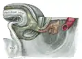 Tail end of human embryo thirty-two to thirty-three days old.
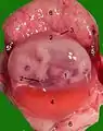 Opened uterus with cat fetus in midgestation: 1 umbilicus, 2 amnion, 3 allantois, 4 Yolk sac, 5 developing marginal hematoma, 6 maternal part of placenta (endometrium)
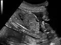 Ultrasound of fetus showing urachus duct from bladder to the umbilicus.
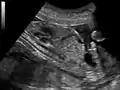 Ultrasound of fetus showing urachus duct from bladder to the umbilicus.
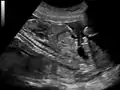 Ultrasound of fetus showing urachus duct from bladder to the umbilicus.
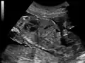 Ultrasound of fetus showing urachus duct from bladder to the umbilicus.
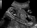 Ultrasound of fetus showing urachus duct from bladder to the umbilicus.
参考文献
- Downs, K.M. 1998.
- First AID for the USMLE Step 1 2008
- http://www.answers.com/topic/allantois

This article is issued from Wikipedia. The text is licensed under Creative Commons - Attribution - Sharealike. Additional terms may apply for the media files.