肛門挙筋
ギャラリー
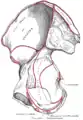 Right hip bone. Internal surface.
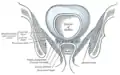 Coronal section of pelvis, showing arrangement of fasciæ. Viewed from behind.
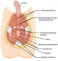 Muscles of male perineum.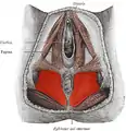
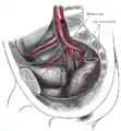 The arteries of the pelvis.
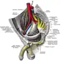 Sacral plexus of the right side.
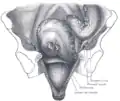 Iliac colon, sigmoid or pelvic colon, and rectum seen from the front, after removal of pubic bones and bladder.
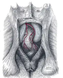 The posterior aspect of the rectum exposed by removing the lower part of the sacrum and the coccyx.
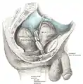 Male pelvic organs seen from right side.
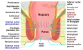 Anatomy of the human anus.
出典
外部リンク
- 『肛門挙筋』 - コトバンク
- Anatomy figure: 41:05-00 at Human Anatomy Online, SUNY Downstate Medical Center —「女性の浅会陰隙の筋肉」。
- Anatomy figure: 42:04-00 at Human Anatomy Online, SUNY Downstate Medical Center —「男性の浅会陰隙の筋肉」。
- Anatomy photo:43:16-0102 at the SUNY Downstate Medical Center —「骨盤横隔膜の筋肉」
- Anatomy image:9072 at the SUNY Downstate Medical Center
- Anatomy image:9089 at the SUNY Downstate Medical Center
- Anatomy image:9871 at the SUNY Downstate Medical Center
- Cross section image: pelvis/pelvis-e12-15ウィーン医科大学のプラスティネーション研究所
- perineum at The Anatomy Lesson by Wesley Norman (Georgetown University)( analtriangle3 )
- pelvis at The Anatomy Lesson by Wesley Norman (Georgetown University)( femalepelvicdiaphragm 、 malepelvicdiaphragm )
- 肛門挙筋症候群に関するメルクのマニュアル記事


This article is issued from Wikipedia. The text is licensed under Creative Commons - Attribution - Sharealike. Additional terms may apply for the media files.